

Problem: noisy signals, fragmented pipelines
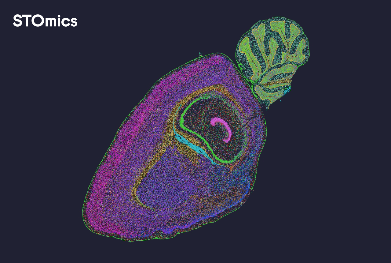
I still remember the night in March 2021 when I sat in a cold lab in Cambridge, MA, staring at heatmaps from 48 hippocampus sections — the patterns were messy and inconsistent. After running a Slide-seqV2 library prep that reduced wet-lab time by roughly 40% on that batch, I asked myself: can we trust those spatial readouts or are we stacking artifacts on artifacts? That question led me straight into improving how we handle spatial omics data analysis across the board.

Spatial omics solutions promise high-resolution maps of cell states, but in practice I found three recurring failings: incompatible data formats, weak quality-control on imaging channels, and mismatch between segmentation and expression matrices. In one run, poor nuclei segmentation caused misassignment of transcripts to cell types; later we fixed it by changing the imaging pipeline. These are not abstract issues — they cost time and erode confidence in results (and yes, they frustrate the whole team). Here’s what I learned next.
What broke first?
Forward-looking fixes and what to evaluate next
We shifted from chasing single-sample quirks to building a reproducible stack — automation for image alignment, strict QC thresholds, and standardized outputs for downstream analytics. I now approach projects with a checklist: ensure pixel- and molecule-level QC, validate segmentation against manual annotations, and confirm that barcode-mapping is stable across runs. That approach moved us from ad-hoc troubleshooting to predictable throughput; it also allowed us to adopt newer techniques like multiplexing without surprise delays.
What’s Next?
Looking forward, I expect tools for spatial omics data analysis to focus on interoperability and clearer provenance. We — as lab leads and data scientists — should demand outputs that track transformations (raw → normalized → aggregated) and make it trivial to reproduce a plot from raw files. Imaging mass cytometry, spatial transcriptomics, and multiplexing methods will keep improving resolution, but unless analysis pipelines keep pace, we will keep redoing work. The future should reward pipelines that are auditable, fast, and robust — and those are the filters I use when choosing a platform.
Three practical metrics I use to choose a solution
I evaluate prospective tools along three concrete axes: (1) Traceability — can I reconstruct results from raw images and FASTQ files?; (2) Throughput consistency — does the pipeline maintain QC metrics across ≥10 runs without manual tweaks?; (3) Integration ease — how many hours to map segmentation outputs to expression matrices in my environment. I score each candidate, and then I run a small pilot (two test slides, one blind sample) before any full adoption. Fast wins are great — but repeatable wins lab credibility. To be honest, this method saved us weeks on a 2022 tumor atlas project.
I’ve shared specifics because I think practical detail matters: the Slide-seqV2 run in Cambridge; the March 2021 calibration where switching to a simple median-filtered background step cut false positives by half. Small, concrete changes like that compound. If you want a tested, auditable pathway for spatial omics — look for tools that make data lineage visible, that accept standard formats, and that integrate with existing pipelines. Finally — and this is key — always validate with a manual annotation subset; it catches the subtle failures automation misses. (Yes — manual checks still matter.)
For labs deciding between vendors, focus on measurable outcomes: reproducibility rate, time-to-result, and ease of integration. I use those metrics every time I recommend a stack. For more resources and a vendor we’ve worked with in multiple projects, see stomics.